
Uterus
Organ for embryonic and fetal development in female mammals.

DeuxPlusQuatre · CC0
The uterus (from Latin uterus, pl.: uteri or uteruses) or womb is the organ in the reproductive system of most female mammals, including humans, that accommodates the embryonic and fetal development of one or more fertilized eggs until birth. It is a hormone-responsive sex organ that contains glands in its lining that secrete uterine milk for embryonic nourishment. The term uterus is also applied to analogous structures in some non-mammalian animals.
- location
- Pelvic region, behind and overlying the bladder, in front of the sigmoid colon
- size
- About 7.6 cm long, 4.5 cm broad, 3.0 cm thick
- weight
- About 60 grams in a typical adult
- layers
- Endometrium, myometrium, perimetrium
- primary function
- Prepare for implantation of a zygote and maintain pregnancy
- position
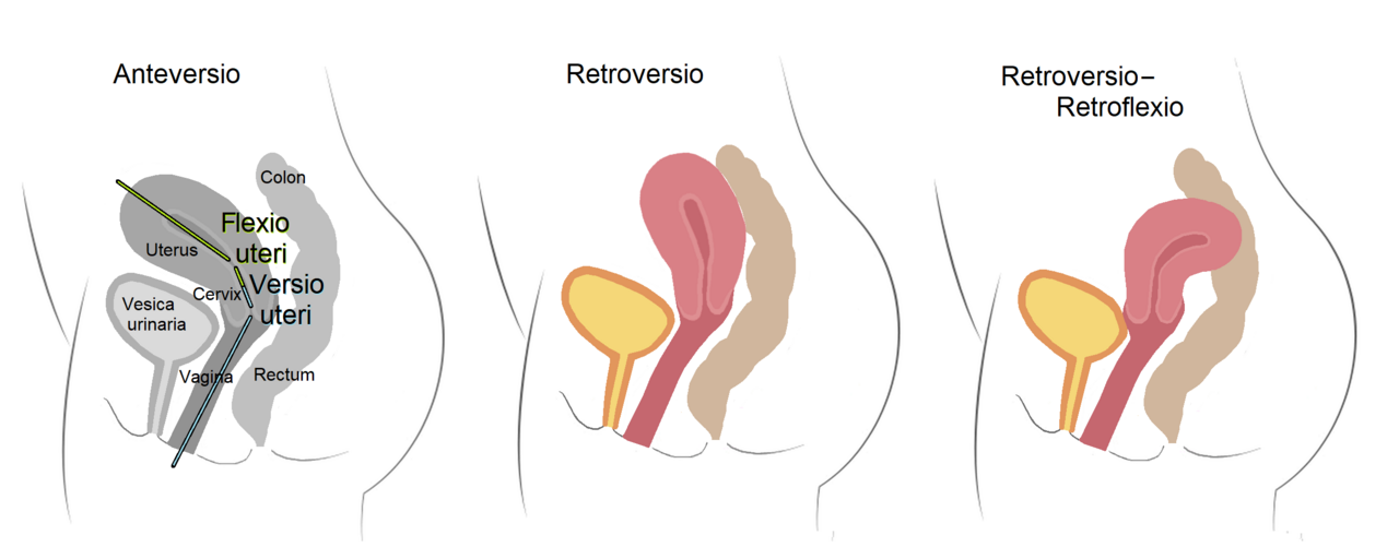
- Anteverted in 50% of women, retroverted in 25%, midposed in 25%
Lore & Background
In humans, the uterus is pear-shaped and located within the pelvic region immediately behind and almost overlying the bladder, and in front of the sigmoid colon. It can be divided anatomically into four regions: the fundus, body, cervix, and cervical canal. The uterus is held in position by ligaments including the pubocervical, cardinal, and uterosacral ligaments, and is covered by the broad ligament of peritoneum. The uterine wall has three layers: the inner endometrium, the middle myometrium of smooth muscle, and the outer perimetrium. The endometrium has a basal and functional layer; the functional layer thickens and is shed during the menstrual cycle or estrous cycle.
Reader's Guide
The uterus is central to mammalian reproduction, providing the environment for embryonic and fetal development. In humans, it prepares for implantation of a fertilized egg and sustains pregnancy through the formation of the placenta. The organ's structure and position vary among mammals, with some having a simplex uterus (as in humans) and others a duplex uterus. The uterus is supported by the pelvic diaphragm, perineal body, and urogenital diaphragm, and its position can be anteverted, retroverted, or midposed. Clinical significance includes assessment of fetal growth via fundal height and various pathological states such as retroversion, prolapse, and congenital malformations like didelphic or bicornate uterus. The term 'uterus' is consistently used in medicine, while 'womb' is common in everyday contexts.
Did You Know?
- The uterus develops from the paramesonephric ducts, which fuse into a single organ known as a simplex uterus in humans.
- The uterus is supplied by arterial blood from the uterine artery and the ovarian artery.
- In the absence of pregnancy, withdrawal of estrogen and progesterone triggers shedding of the functional layer of the endometrium, resulting in menstruation.
- The uterus is mobile and moves posteriorly under pressure of a full bladder or anteriorly under pressure of a full rectum.
The Uterus as the Stage of Life
The uterus occupies a central role in the journey from a single fertilized cell to a living newborn. While the initial meeting of sperm and egg most commonly takes place within the fallopian tubes, fertilization can also occur inside the uterus itself. Once the zygote forms, it travels to and implants in the uterine lining, where the remarkable processes of embryogenesis and morphogenesis unfold over the course of a typical thirty-eight-week gestation. The uterus provides the environment in which the embryo matures into a fetus capable of independent life. When that readiness arrives, the cervix dilates and powerful uterine contractions drive the infant through the birth canal—the vagina—into the external world. This final act, known as childbirth, marks the transition from internal to external existence. The entire sequence, from implantation to delivery, positions the uterus not merely as a passive container but as an active, muscular participant in sustaining and ultimately releasing new human life.
Anatomical Partners in the Female System
The uterus does not operate in isolation; it is one component of a broader female reproductive architecture that includes both external and internal structures. The external genitalia, collectively called the vulva, serve as the entry point, while the internal system encompasses the ovaries, fallopian tubes, uterus, and cervix. The female sex cells—ova—are notably larger than their male counterparts and are formed within the ovaries during fetal development, long before a woman is born. They carry nutrients destined for the future zygote and embryo. On a regular hormonal schedule known as the menstrual cycle, one ovum matures through oogenesis and is released into the fallopian tube. If no fertilization occurs, the unfertilized egg is expelled from the body through menstruation. The ova themselves reside in a dormant, prophase-arrested state within primordial follicles, yet they retain the capacity for highly efficient DNA repair, safeguarding the genetic integrity that will one day be passed to offspring.
The Gametes and the Odds of Conception
Conception is a statistical marvel set against enormous odds. A typical ejaculation carries roughly two hundred and fifty million sperm cells, yet only about one in fourteen million will successfully navigate the vagina and cervix to reach the fallopian tube. Each gamete—sperm and ovum alike—carries just half the forty-six chromosomes found in ordinary body cells, and their merger into a single zygote is the moment genetic recombination takes place. The sperm that ultimately fertilizes the egg does so with the aid of hydrolytic enzymes housed in its acrosome. Both male and female germ cells possess specialized DNA repair mechanisms: homologous recombinational repair and non-homologous end joining in the testes, and efficient homologous recombinational repair in the oocytes. These processes mend double-strand breaks and other damage, ensuring that the genome handed to the next generation remains as intact as possible.
Beyond the Biology — Law, Medicine, and Society
Human reproduction is shaped by forces well beyond cellular biology. Before any reproductive act can occur, both parties must have passed through puberty—ovulation in females and the first ejaculation in males—since pre-pubertal genitalia lack reproductive function. Legal frameworks add another layer: a person below the age of consent cannot legally authorize sexual intercourse or artificial reproductive procedures, and violations can trigger charges of statutory rape or child sexual abuse depending on jurisdiction. Even among consenting adolescents, comprehensive sex education emphasizes contraception to prevent both sexually transmitted infections and unplanned pregnancy. In developed nations, adolescent pregnancies are particularly discouraged because of elevated maternal and infant mortality risks tied to the body's incomplete physical readiness. On the medical front, assisted reproductive technologies such as in vitro fertilization and artificial insemination offer pathways to conception that bypass sexual intercourse entirely, expanding the definition of how new life can begin.
Gallery


Frequently Asked Questions
What is the uterus?
The uterus, also called the womb, is a hormone-responsive reproductive organ found in most female mammals, including humans. It serves as the site where a fertilized egg implants and where embryonic and fetal development takes place until birth.
Where exactly is the uterus positioned in the body?
It sits in the pelvic region, tucked behind and above the bladder and in front of the sigmoid colon. In terms of tilt, roughly half of all women have an anteverted uterus, a quarter are retroverted, and the remaining quarter fall into a midposed position.
What are the three layers of the uterine wall?
The uterine wall is built from three distinct layers: the inner endometrium, the thick muscular middle layer called the myometrium, and the outer serous layer known as the perimetrium. The endometrium also houses glands that produce uterine milk to nourish an early embryo.
How large and heavy is a typical adult uterus?
In a typical adult, the organ measures roughly 7.6 cm in length, 4.5 cm in width, and 3.0 cm in thickness, weighing around 60 grams. These dimensions can shift dramatically during pregnancy as the organ expands to accommodate a growing fetus.
What is the primary job of the uterus in reproduction?
Its core role is to prepare the internal lining for zygote implantation and then sustain the pregnancy through to delivery. The term is also applied to analogous structures in certain non-mammalian species, showing how widespread this developmental function is across animal groups.
More in Reproduction & Development 1-19
Spotted an error? Know more?
This is a living reference — every entry is fact-audited, and reader corrections feed straight into our audit queue. Suggest an edit · See this site's audit record